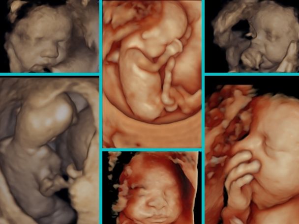
There are 2D, 3D and 4D ultrasounds. All ultrasound uses sound waves to create an image of the unborn baby on the screen. A 2D ultrasound of a fetus creates a black-and-white image. The main scan is done in 2D only. We take images of the unborn baby in grey scale. We take measurements of the unborn baby, assess its growth and assess the structure of the baby mainly in 2D.
Nowadays, 3D- three dimensional and 4D - fourth dimensional ultrasounds have become common. Usually, all ultrasound machines have software and volume probes with which we do 3D/ 4D scan.
3D scan makes three dimensional images of the unborn baby like that of a face. When we see these images in real time, that is, we see the movements of the unborn baby as well, it is called 4D ultrasound. Some centres have started adding 5D ultrasound also. 5D ultrasound is nothing but an extension of 3D ultrasound only.
The main scan is done in grey scale, that is 2D scanning. 3D and 4D ultrasounds can be used by the person to add more information to it but are not absolutely necessary.
What laymen understand is that , by means of 3D and 4D ultrasound , the parents can see realistic images of the face of the baby. It adds to great experience in pregnancy. But it is not limited to taking pictures of baby’s faces.
The benefits of 3D and 4D ultrasound are limited. It becomes useful when any structural abnormality is suspected. It helps us to take views of the baby from different angles and provides more depth to our understanding of abnormalities. 3D and 4D ultrasounds can be done to add more details which helps us in counselling couples. Like when we see a gap in the lips- cleft lip, 3d and 4D ultrasound helps to know the extent of the gap, whether there is involvement of palate also.
3D and 4D ultrasounds are not generally part of daily prenatal tests. What is interesting is that 3D ultrasounds provide the three-dimensional image of your infant, while 4D ultrasounds provide a live visual effect, such as a clip, you can see your baby smile or yawn.
It is done from your tummy. A gel is placed on your tummy and then a probe is moved over the tummy to do the ultrasound. It can also be done vaginally but usually done in early pregnancies.
Looking at the face of the baby during pregnancy increases the bonding of parents with the baby. But sometimes, it is not possible to provide you with a good image. It depends on how the baby is lying or if you have a lot of tummy fat. So, discuss it with your ultrasonologist.
Ultrasound uses sound waves to create an image. Till now, no significant deleterious effects of ultrasound have been seen on developing babies inside the mother's womb. 3D and 4D ultrasound also uses the same sound waves to create images, hence, no harmful effects on the unborn baby.