-
Email fetalmedicinesolutions@gmail.com
-
Phone +91 9911 411 982
-
Office Hours Mon - Sat: 9AM - 6PM
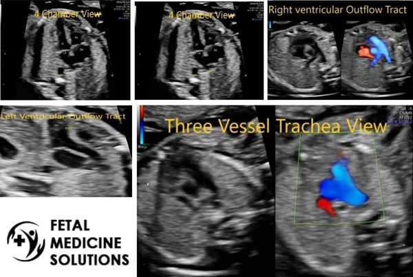
Fetal echocardiography is nothing but a detailed ultrasound the fetal heart. The heart and its connections, blood flows and their velocities are checked to identify and characterize the fetal heart anomalies
Fetal echocardiography is only about fetal heart evaluation whereas in an anomaly scan, besides heart, other structures are also assessed. Normally, fetal heart assessment requires minimum evaluation of 4 chamber view, outflow tracts and 3 vessel views.
Many people restrict the assessment to 4 chamber view and outflow tracts and some only to 4 chamber view. This is not enough to rule out cardiac abnormalities. If these are not assessed in the anomaly scan, then fetal echocardiography is advised. We assess the fetal heart in detail in anomaly scan only.
Fetal echocardiography is indicated if there is suspicion of cardiac structural or functional abnormality, abnormal fetal heart rate or rhythm, multiple pregnancy, increased nuchal translucency (fluid behind neck of the baby), extra-cardiac abnormality or if mother has diabetes or auto-immune disease, history of heart disease in parents, siblings or previous pregnancy. Obesity or suboptimal views or suboptimal assessment during anomaly scan can also warrant fetal echocardiography.
No, we cannot. There are certain lesions like aortic stenosis, coarctation of aorta which might get apparent later as they evolve over time. However, the aim is to maximise the probability of detecting and correctly identifying clinically relevant congenital heart diseases.
It is usually done between 18-22 weeks, although some structure might be better identified before or after this period. It is possible to assess the fetal heart early like at the time of first trimester screening scan when certain heart defects can be recognised or atleast a suspicion can be raised.
We follow the protocol as laid down by ISUOG (International Society of Ultrasound in Obstetrics and Gynaecology) 2013 and AIUM (American Institute of Ultrasound Medicine) 2020.
Ultrasonography is absolutely safe for clinical practice. To date, there has been no independently confirmed study to suggest otherwise.