-
Email fetalmedicinesolutions@gmail.com
-
Phone +91 9911 411 982
-
Office Hours Mon - Sat: 9AM - 6PM
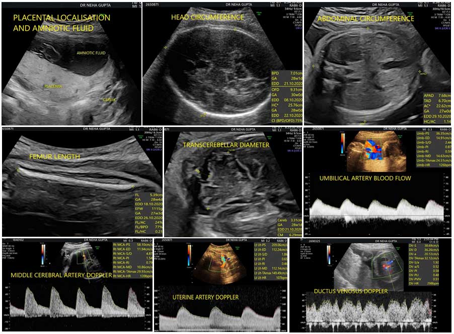
This scan is meant to assess the fetal weight, the fluid around the baby, the placental localisation and the blood flows to the baby.
Fetal dopplers mean assessment of baby’s blood flows. Maternal blood flows to the uterus are assessed by uterine artery Dopplers. Blood flow through cord is called as umbilical artery blood flow. Blood through baby’s brain is called as middle cerebral artery Dopplers and through tummy is referred to as ductus venosus Dopplers.
The aim is to assess the fetal growth disorders and the well being of the unborn baby. If the growth is appropriate for the weeks, then it is normal. If the growth is small, then it is a weak baby, also called as fetal growth restriction. If the growth is high, then it is a big baby, also called as macrosomia.
Besides fetal weight, maternal history and symptoms, fluid assessment and baby’s blood flows helps us to further identify the babies at risk of adverse outcome. Depending on the above factors, appropriately delivery can be timed.
No, minimum two growth scans are required to assess the fetal well being. One growth scan gives us the fetal growth assessment at a given time. However, two helps us to establish the growth velocity, and identify the at risk fetuses.
A detailed structural assessment is difficult after 26-28 weeks due to fetal position and baby does not move around completely as it does around 20 weeks. But still, whatever is possible due to fetal position and advanced gestational age, is assessed at the time of growth scans.
Yes, it is possible. The risk of detection of structural abnormality in the third trimester is 1 in 200 pregnancies. These could be those that develop later on like certain brain abnormalities, kidney problems or those which were missed at the time of anomaly scan.
Usually, the growth and Doppler assessment start from 28 weeks onwards. Most of the times, two scans are done for the growth assessment- one around 28 weeks and other between 34-37 weeks. Sometimes, depending on the growth and blood flows or specific history, more scans may also be required. This will be explained to you by the fetal medicine specialist.
No. But at this time, if the fetal position is appropriate, good 3D and 4D views can be obtained of the face of the fetus. We routinely take 3D/4D views of the face if the baby’s position is appropriate.
Individuals who routinely perform obstetric scans and have the necessary requirements as per the law of the land.
Prenatal ultrasonography appears to be safe for clinical practice. To date, there has been no independently confirmed study to suggest otherwise.